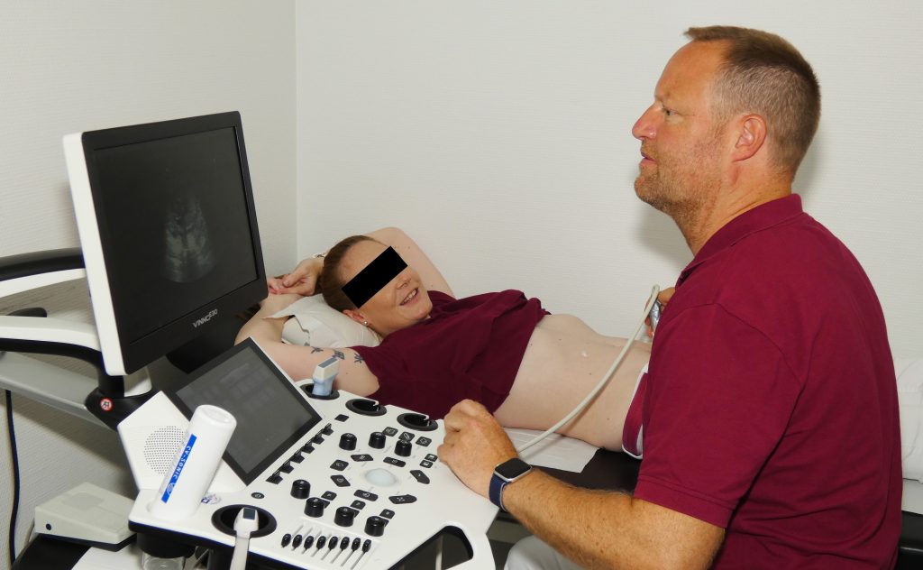
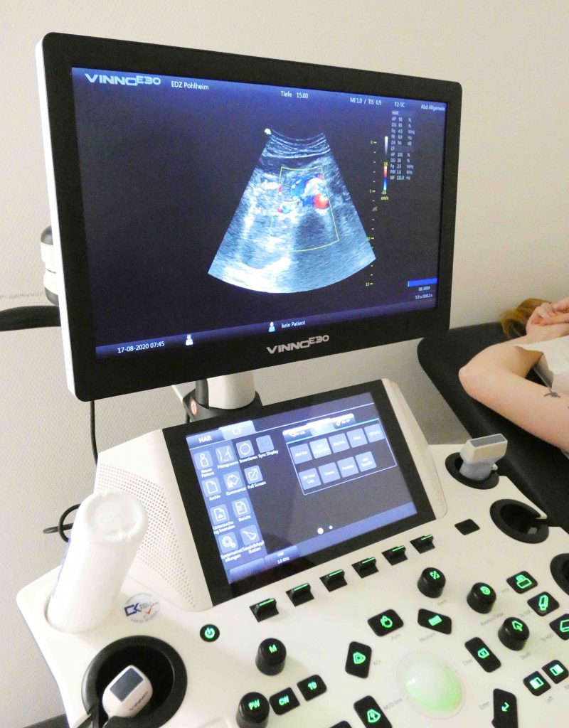
The presence of an accessory pathway, which extends unidirectionally from the ventricle to the atrium, but not in the opposite direction, is not apparent from the analysis of the ECG during the sinus rhythm, since the ventricle is not pre-excited (Fig. 37.12). Therefore, electrocardiographic manifestations of WPW syndrome are absent and the accessory pathway is „hidden“. Since the mechanism responsible for most TAs in patients with WPW syndrome is macroreintegration caused by anterograde conduction through the AV node – its beam path and retrograde conduction via an accessory pathway, even if it is only performed retrograde, the accessory pathway, even if it is performed only retrograde, can still participate in the retransmitted circulation and cause reciprocal TACHYCARDIA AV. During electrocardiographic examination, tachycardia resulting from this mechanism may be suspected if the QRS complex is normal and the retrograde P wave occurs after completion of the QRS complex, in the ST segment or at the beginning of the T-wave (see Fig. 37.8C). Sometimes the P wave is not clearly visible and can lead to ST segment depression; If this is observed during TA, the mechanism of arrhythmia is most often a re-entry with an accessory pathway (AV reentrant tachycardia). In addition, in this context, ST depression, which occurs only during TA (disappears with the cessation of tachycardia), does not indicate ischemia if there are no other signs of ischemia (chest pain, enzymatic increase, known coronary artery disease). One caveat to the statements just made is that if the His beam catheter is perpendicular to the underlying beam, directional information may be lost. If a more sensitive mapping of its beam is desired, a higher density mapping electrode (with narrower electrodes) may be required. Electrocardiogram of a patient with pulmonary embolism with sinus tachycardia of about 150 beats per minute and right trunk block.

Contribution from Wikimedia Commons, Walter Serra, Giuseppe de Iaco, Claudio Reverberi and Tiziano Gherli (more…) The last two ventricular beats with a cycle duration of 600 milliseconds are represented in A. A premature stimulus (S2) at the 260 millisecond range of S1-S2 and another premature stimulus (S3) at a cycle length of 210 milliseconds initiate a sustained monomorphic VT at a cycle length of 300 milliseconds. B, Two premature ventricular stimuli (S1-S2) produce an unstable VT that lasts several beats at a shorter cycle time (230 milliseconds) and then terminates, followed by a sinus rhythm. HBE, its beam electrogram; RV, right ventricle. Background: Current definitions of LBBB cannot always distinguish LBBB from left ventricular conduction delay. Only patients with LBBB should normalize to its beam rate. Patients who develop a new LBBB immediately after transcatheter aortic valve replacement (TAVR) provide an excellent model for defining the characteristics of the electrocardiogram (ECG) of the LBB. We tried to describe their ECG characteristics and develop a new ECG definition of LBBB. The His bundle is a group of fibers that carry electrical impulses through the center of the heart. If these signals are blocked, you will have problems with your heart rate.

A complete AV block can result from a block at the AV node (usually congenital;Fig. 40.10), in the bundle of His or distal to it in the Purkinje system (usually acquired;eFig. 40.8).2 Proximal block to the His bundle usually has normal QRS complexes and rates of 40 to 60 beats / min, since the focus escapes that controls the ventricle, in or near Its packet. In the complete AV node block, the P wave is not followed by a Deviation of Sound, but each ventricular complex is preceded by a Deviation of Sound (Fig. 40.10). Its beam recording may be useful for distinguishing AV-Nodal from the intrahisian block, as the intrahisian may carry a more severe prognosis than the AV node block. Intrahisian block is rarely detected without invasive studies. In patients with AV lymph node block, atropine usually accelerates atrial and ventricular levels. Exercise can reduce the extent of AV node blockage. Acquired complete AV block most often occurs distally at the His bundle due to trifascicular conduction disorder.

Each P wave is followed by a deviation of the being, and the ventricular exhaust complexes are not preceded by a deviation of the being (see Fig. 40.8). The QRS complex is abnormal and the ventricular level is usually less than 40 beats/min. An inherited form caused by the degeneration of the branches of the His bundle and the beam has been linked to the SCN5A gene, which is also responsible for LQT3 (see Chapter 33). A third-degree AV block or a full AV block occurs when no atrial activity is directed to the ventricles, and therefore the atria and ventricles are controlled by independent pacemakers. Thus, the complete AV block is a type of complete AV dissociation. The atrial pacemaker may be a sinus or fallopian tubes (tachycardia, floating or fibrillation) or may result from a FOCI of AV crossing occurring above the block with retrograde atrial conduction. The ventricular focus is usually just below the block area, which may be above or below the bifurcation of the His beam.

Ventricular pacemaker activity sites located in or closer to the His bundle appear to be more stable and may produce a faster escape rate than those further into the ventricular conduction system. The ventricular rate in the acquired complete heart block is less than 40 beats/min, but may be faster with a congenital full AV block. The ventricular rhythm, which is usually regular, can vary in response to premature ventricular complexes (PVCs), movement of the pacemaker site, irregular discharge from the pacemaker focus, or autonomic influences. The electrogram of His beam (Being) represents the recording of a potential of Being, and the ventricular electrogram (V) represents the activation of the ventricles. When the branch of the right beam is interrupted, electrical stimuli are conducted from the atrioventricular (AV) node to the His beam and the left beam branches down. The left ventricle depolarizes first, while the right ventricle later polarizes, leading to the characteristic results of the ECG. [13] Conclusion: We have developed a new ECG definition of LBBB, which contains 2 new discoveries: the notching/washing of the R wave in at least one lateral line and an R wave ≤20 ms in V1. Further larger studies are warranted to confirm these findings. Its beam electrography is a test that measures electrical activity in a part of the core known as the His beam. The His bundle is a group of fibers that carry electrical impulses through the center of the heart to ensure that the heart is beating properly.

His beam electrography is part of an electrophysiological (EP) study. An intravenous catheter (IV line) is started in your arm to allow medication to be administered during the procedure. Electrocardiogram (ECG) lines are placed on your extremities. A catheter is inserted through a small incision in a vein in your arm, neck or groin, which is carefully threaded into the heart using an X-ray imaging technique called fluoroscopy to guide the insertion. The ECG monitors your heart for arrhythmias (abnormal heart rhythms) during catheter placement. The catheter, which is equipped with an electrode, then measures the electrical activity of the His beam. A right beam branch block is usually caused by pathological processes that modify the myocardium of the right beam branch, for example, structural changes, trauma and infiltration processes. In rare cases, hyperkalemia can alter the physiology of conduction by slowing down the conduction of electrical impulses through heart tissue, resulting in blockage of the right beam branch. [6] Infections such as myocarditis or myocardial infarction can cause direct cell damage to the right branch of the trunk. [2] Increased right intraventricular pressure, acute by pulmonary embolism or chronic as in the pulmonal horn, can stretch the right branch of the bundle, resulting in blockage of the beam branch. [7] RbBB can also be induced by iatrogene from straight cardiac catheterizations and by ethanol ablation for septal reduction of hypertrophic cardiomyopathy.

[3] [8] Idiopathic fibrosis and calcification of the conduction system, called Lenegre`s disease or Lev`s disease, are a less common cause of right beam branch block, but are more common in the elderly. [9] [10] A tachycardia-dependent bundle branch block may occur in patients with underlying heart disease that causes degeneration of the conduction pathway.